Anterior Interosseous Nerve Repair
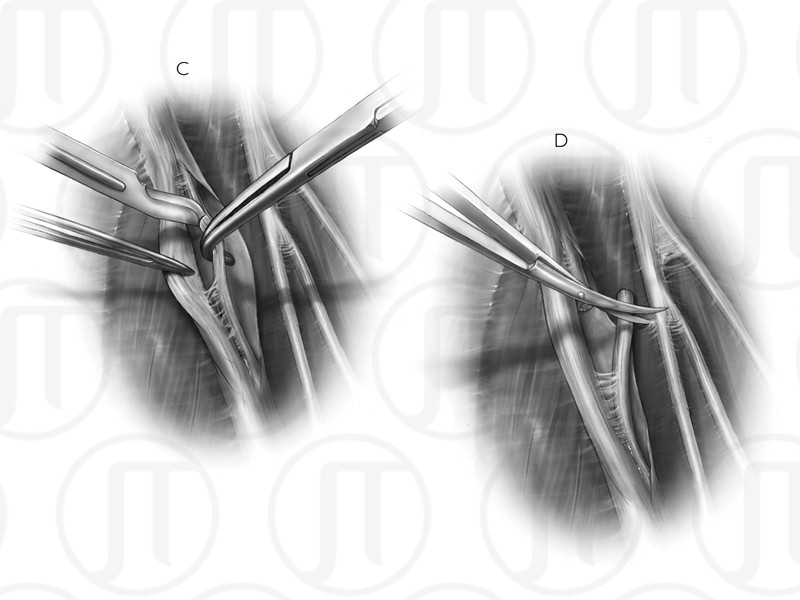
The surgeon first isolates and test the damaged nerve (Figure 01). The non-functioning and functioning nerves are cut (Figure 02). The nerves are then trimmed and adhered using fibrin glue (Figure 03).
Anatomy in this medical illustration: Lateral cutaneous nerve, Musculocutaneous nerve, Median nerve, Anterior interosseous fasicle of median nerve, Brachialis muscle


